SSR Inst. Int. J. Life Sci., 9(1):
3136-3140,
January 20223
Dystocia Due to
Secondary Uterine Inertia and its Obstetrical Management- A Case Report
Hima Bindu Kolagani1*, Chandra Prasad Borra2, Srinivas Manda≥
1PG
Scholar, Department of Veterinary Gynaecology and
Obstetrics, NTR College of Veterinary Science, Gannavaram,
Sri Venkateswara Veterinary University, Tirupati, India
2Assistant
Professor, Department of Veterinary Clinical Complex, NTR College of Veterinary
Science, Gannavaram, Sri Venkateswara Veterinary
University, Tirupati, India
3Professor,
Department of Veterinary Gynaecology and Obstetrics,
NTR College of Veterinary Science, Gannavaram, Sri
Venkateswara Veterinary University, Tirupati, India
*Address for Correspondence: Dr.
Hima Bindu Kolagani, PG Scholar, Department of
Veterinary Gynaecology and Obstetrics, NTR College of
Veterinary Science Gannavaram, Sri Venkateswara
Veterinary University, Tirupati, India
E-mail: bindukolagani@gmail.com †
ABSTRACT- Background: A full-term pregnant Ongole cow was
presented to the Large Animal Obstetrical Ward with a history of reduced feed
intake, dull and ruptured foetal membranes 24 hours
before presentation without progress in parturition. The temperature was within
the normal physiological range. Per-vaginal examination revealed second-degree
cervical dilation, lack of uterine and abdominal contractions and the vaginal
discharges were reddish brown and putrid.
Methods: The case was diagnosed as secondary uterine inertia and treated
with an intracervical application of misoprostol and intravenous calcium
therapy.
Results: Three
hours
after the application of misoprostol and calcium therapy, full dilatation of
the cervix was achieved to facilitate the delivery of the dead male emphysematous
foetus by traction. Uneventful recovery of the dam
was noticed.
Conclusion: Usage of misoprostol along with CMC massage and calcium therapy
resulted in speedy recovery of dystocia suffering with incomplete cervical
dilation.
Key Words: Ongole cow, Emphysematous foetus, Maternal Dystocia, Misoprostol, Carboxy methyl
cellulase, Secondary uterine inertia
INTRODUCTION-
Dystocia is a common obstetrical problem that occurs when the first or second
stage of labour was prolonged and delivery requires
assistance [1]. primarily the case history should be taken since it
is necessary for the handling of dystocia [2]. For assessing an
individual animal and formulating a clinical management plan, it is advised to
divide the causes of dystocia into those of foetal
origin or maternal origin [3].
The causes may be maternal (uterine inertia,
inadequate size of birth canal) or foetal factors
(oversized foetus, abnormal orientation of foetus) [4]. Incomplete cervical dilation of
maternal causes is found to be a more frequent cause of dystocia [5].
Dystocia due to secondary uterine inertia obstructs to delivery of the dead foetus through the cervical canal and is rarely observed in
companion animals [6]. Secondary uterine inertia commonly occurs due
to uterine exhaustion leading to obstructive dystocia [7]. The
condition occurs mainly due to an obstruction in the birth canal or can happen
spontaneously at the second stage of parturition [8]. Weak uterine
contractions often resulted in relatively inefficient dilation of the ripened
cervix, which is the probable pathogenesis.†††
The
period, when the cervix is fully dilated is relative to short duration. If the
calf is not delivered through the ripened cervix, it will start to involute,
thus trapping the foetus within the uterus [9].
CASE PRESENTATION- A full-term pregnant Ongole cow in its fourth parity was presented to the Large
Animal Obstetrical Ward with a history of ruptured foetal
membranes 24 hours before presentation without progress in parturition.
Clinical examination revealed relaxation of sacro-sciatic
ligaments, enlarged udder and edematous vulval lips. Per rectal examination
revealed the absence of fremitus, foetal movements,
and foetal reflexes with crepitation. Upon
obstetrical examination revealed second-degree cervical dilation, and lack of
uterine and abdominal contractions; the birth canal was dry and inflamed with
reddish brown and putrid vaginal discharges. The presentation, position and
posture of the dead emphysematous foetus could not be
fully determined as the cervix was not fully dilated. The case was handled by a
field veterinarian, who failed to deliver and thus, referred the case.
The animal was restrained
by caudal epidural anaesthesia @ 5 ml of 2% lignocaine hydrochloride solution.
The animal was treated with Inj. Valethamate bromide
(Epidosin 50 mg IM), Inj. Dexamethasone sodium
phosphate (Dexacare-Vet 20 mg IM), Inj. Cloprostinol sodium (Pragma 500 Ķg IM) along with fluid
therapy (25% Dextrose and Ringers Lactate @ 2 litres
each) for rehydration. Feathering of the cervix was done by massaging the cervix
with arms by using lukewarm carboxy methyl cellulose (CMC) for 15 minutes.
After 1 hour of feathering slight progress in cervical dilatation was noticed
but was not sufficient to facilitate the delivery of the dead foetus. As the texture of the cervix was moderately soft
and partially lobulated, it was decided to dilate the cervix's medical
management. Carboxymethyl cellulose gel mixed with Tab. Misoprostol @ 1600 mcg
(Misoprost 200 mcg, Cipla, 8 tabs) was applied
intra-cervically. Three hours after the application of Misoprostol, complete
dilatation of the cervix was achieved. Detailed obstetrical examination
revealed the presence of a dead emphysematous foetus
in anterior longitudinal presentation, dorso-sacral
position with head, neck and forelimbs extended into the birth canal. A slow
intravenous infusion of 150 ml calcium borogluconate
was administered to improve the uterine tone. Carboxymethyl cellulose solution
(2%) @ 5-6 litres was infused into the uterus as a
lubricant. Calving ropes were applied above the fetlock joints of both the
forelimbs and the loop of the snare was applied to the neck behind the ears. As
the foetus was dead and emphysematous, the gas from
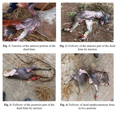
the sub-cutaneous tissues was removed by incising the dorsal areas of the foetus with a blunt scalpel (Fig. 1). Forced traction led
to detachment of the posterior portion of the foetus
and only the anterior portion of the foetal body
could be delivered by traction (Fig. 2). Hindlimbs of the foetus
were identified and version was performed on the posterior portion of the foetus to extend the hindlimbs into the birth canal. The
loop of the calving ropes was placed above the fetlocks of both hindlimbs and
the posterior portion was delivered by traction (Fig. 3). Further, some
the internal organs of the foetus like kidney, liver
and intestines were removed in portions. After removal of foetus
and its parts (Fig. 4), the uterine lumen was thoroughly examined to rule out
the presence of tears and haemorrhage.

Animal was treated with
Inj. Oxytocin 25 IU as single dose, Inj. Moxel 3 gms
BID (Amoxycillin sodium 1500 mg and Cloxacillin sodium 1500 mg), Inj. Megludyne @ 2.2 mg /kg body weight (Inj. Flunixin
meglumine), Inj. Zeet 60 mg (Inj. Chlorpheniramine
maleate) and fluid therapy with 2 litres of Ringers
lactate and Normal saline, each. Antibiotic therapy was continued for 5 days
post-operatively and the cow experienced an uneventful recovery.
DISCUSSION- All obstetrical cases
were to be treated as emergencies [10]. Incidence of dystocia can be
prevented by good nutrition, and by reducing the fetomaternal
disproportions by appropriate dam and sire selection [11].
Incomplete cervical dilation may occur both in the multiparous animals and
heifers. Dystocia caused exhaustion of the uterus leading to
secondary uterine inertia and the affected cows had a very low survival rate [12].
Obstruction
or a maldisposed or oversized fetus in the birth
canal could result in dystocia, which if not corrected in time lead to uterine
muscle exhaustion and resulted in a condition called secondary uterine inertia [13].
The condition was characterized by the presence of weak and transient uterine
contractions with the inability to deliver the foetus.
The onset of parturition was normal with rupture of the foetal
membranes, but the long duration of the dystocia could have led to secondary
uterine inertia and partial involution of the cervix [14].
†In prolonged cases of dystocia, the presence
of a dead foetus at body temperature would cause
putrefaction and bloating of the foetus if delayed
beyond 24-48 hours. Further, prolonged dystocia resulted in weak and
intermittent abdominal contractions within a few hours and later ceased due to
exhaustion. The more prolonged dystocia, the poorer the progress [15].
Ripened
cervix dilation is regulated mainly by myometrial contractions [16].
Weak myometrial contractions can become the causative factor for incomplete
cervical dilation and finally results in dystocia [17]. Secondary
uterine inertia is one of the major factors for incomplete cervical dilation
occurs because of malposition, twin calving, and prolonged dystocia [18].
Prostaglandins play an important role in cervical ripening because of their
increased levels in the fetal membranes, uterus, and cervix [19].
Misoprostol is the analogue of prostaglandin E1, which is commonly used to
ripen the cervix [20].
In the cases of
incomplete cervical dilation, cervical massage with sodium carboxy methyl
cellulose could be done, otherwise leaving the cervix to dilate on its own
causing the hardening of the cervical texture and followed by failure of
dilation [21]. Cervical dilation improved by the combination of the
warm sodium carboxy methyl cellulose gel addition with cervical massage can
induce the activities of collagenolytic enzymes improving cervical softening [22].
cervical massage can be done by warm sodium carboxy methyl cellulose with the
gloved hand at the region of the external os. Calcium
therapy helps in dilating the cervix by improving the uterine contractions
which further resulting the dilation of the cervix [23].
CONCLUSIONS- Maternal factors
especially improper cervical dilation appeared to frequent cause of dystocia
which eventually leads to exhaustion of uterine muscles, causing secondary
uterine inertia. Incomplete cervical dilation is the more frequent cause of
dystocia leading to secondary uterine inertia.
Usage of misoprostol
along with CMC massage and calcium therapy resulted in speedy recovery of
dystocia. Administration of hyaluronidase in the cervix of pregnant animals can
improve cervical ripening and helps to shorten the length of labour without producing any side effects.
Research concept- Manda Srinivas
Research design- Manda Srinivas
Supervision- Borra Chandra
Prasad
Materials- Kolagani Hima Bindu
Data collection- Kolagani Hima Bindu
Data analysis and Interpretation- Kolagani Hima Bindu
Literature search- Kolagani Hima Bindu
Writing article- Kolagani Hima Bindu
Critical review- Borra Chandra
Prasad
Article editing- Borra Chandra
Prasad
Final
approval- Manda Srinivas
REFERENCES
1.
Haben F. Dystocia due to secondary
uterine inertia in Dog and its surgical management. Int J. Clin Stud., 2020;
4(3): 002. Doi:
10.46998/IJCMCR.2020.04.000087.
2.
Kumar P. Applied
Veterinary Gynaecology and Obstetrics. 1st
ed., IBDC International book distributing co: 2009; pp. 132-40.
3.
Youngquist RS, Threlfall
WR.† Current Therapy in Large Animal
theriogenology. 2nd Ed., London; Saunders Elsevier: 2007; pp.
310-33.
4.
Weldeyohanes G. Dystocia in domestic
animals and its management. Int J Pharm Biomed Res., 2020; 7(3): 1-11.
5.
England G. Disorders of
parturition in the Bitch. Veterinary nursing J., 1996; 11(3): 77-84.
6.
Persson G. A survey of
dystocia in the boxer breed. Acta Veterinaria
Scandinavica., 2007; 49(1): 8.
7.
Darvelid AW. Dystocia in the
bitch. J Sma Ani Practi.,
1994; 35(8): 402-07.
8.
Linde-Forsberg C.
Abnormalities in pregnancy, parturition and the periparturient period. Text
Book of veterinary Internal Medicine, 2005; 7: 1890-901.
9.
Noakes DE, Parkinson TJ.
Arthurís Veterinary Reproduction and Obstetrics. 8th ed., London;
Saunders Elsevier, 2013; pp: 229-44.
10. Arthur
GH, Noakes DE, Pearson H. Maternal dystocia treatment. Veterinary Reproduction
and Obstetrics. 8th ed., London; Saunders Elsevier: 2001; pp.
229-44.
11. Dessie
A. Management of dystocia cases in the cattle. J Reprod
Infertility., 2017; 8(1): 1-9.
12. Purohit
GN. Maternal dystocia in cows and buffaloes. Open. J Ani Sci Revi., 2011; 1(2):
41.
13. Jackson
PGG. Dystocia in the Cow. Hand Book of Veterinary Obstertrics.
W. B. Saunders Philadelphia, Philadelphia, 1996; pp: 221.
14. Phogat
JB. Incidence and treatment of various forms of dystocia in buffaloes. Ind J
Ani††† Reprod.,
1992; 13: 69-70.
15. Roberts
SJ. Veterinary Obstetrics & genital diseases. 2nd ed., CBS
publishers and distributors Pvt. Ltd, 2004; pp. 132-40.
16. Lindgren
L. The influence of uterine motility upon cervical dilation in labour. American J Obstet Gynaecol., 1973; 117(4): 530-36.
17. Jitendra
K. Incomplete cervical dilation of cervix in large animal: A review. Pharm Innov J., 2022; 11(2): 573-578.
18. Noakes
DE, Parkinson TJ, England GC. Arthurs veterinary reproduction and obstetrics.
10th ed., London; WB. Saunders Company Ltd: 2018; pp. 277.
19. Fuchs
A, Graddy R. oxytocin induces PGE2 release from bovine cervical mucosa in
vivo. Prostaglandins Other Lipid Mediat., 2002; 70(1): 119-29.
20. Fiala
C, Tang O. cervical priming with misoprostol prior to trans cervical
procedures. Int. J Gynaecol Obstet., 2007; 99:
168-71.
21. Honparich M. cervical massage with
SCMC for achieving complete cervical dilation in successfully detorted uterine
torsion affected buffaloes. Indian J Ani Sci., 2009; 79(1): 26-29.
22. Shi
L, Shi SQ. changes resistance and collagens fluroscence
during gestation in rat. J Perinatol., 1999; 27:
188-94.
23. Shivika
C. Incomplete cervical dilation in large animals: A review. Pharma Innov J., 2022; 11(2): 573-78.