ABSTRACT-
Invasive fungal infections have become a major source of morbidity and mortality in post operative
patients. Critically ill patients after extended surgical procedure are more risk to post surgical fungal infections. Life
saving devices like central venous catheters can increases risk for fungal infections. Surgical infections are infections of
the tissues, organs or spaces exposed by surgeons during performances of surgical procedure. Mold infection is
increasingly common in post operative patients. Postoperative surgical infection represents an uncommon but potentially
devastating complication of surgery. Unfortunately, medical community is not much aware of such secondary infections
due to fungi in post operative patients leading to grave consequences. Better diagnostic methods are needed to improve
the outcome of successful surgery and better health care for public. The diagnosis of invasion and dissemination in the
majority of cases requires the acquisition and proper interpretation of clinical evidence.
Key-Words- Postoperative, Surgical infections, Secondary infections, Diagnostic method
INTRODUCTION-
Fungal pathogenesis is a multifactorial phenomenon.
Pathogenesis is the ability of a microorganism to infect the
host and produce disease resulting from interaction of
pathogen with host via expression of certain factors on both
sides. Pathogenicity of a fungus depends on its ability to
adapt to the tissue environment and to withstand the lytic
activity of the host’s defenses [1]. Postoperative surgical
infection represents an uncommon but potentially
devastating complication of surgery. Mold such as
Aspergillus are the most common opportunistic filamentous
fungi, which can cause a very serious infection that develop
rapidly and that can sometimes be fatal [2]. The infection is
caused by the spores from the environment, which gets into
the body through the lungs, gastrointestinal system or the
skin. Primary infection of the skin may occur only when the skin
is damaged [3-4]. The development of surgical fungal
infection is related to three factors, the degree of microbial
contamination of the wound during surgery, the duration of
the procedure and the host factors, such as diabetes,
malnutrition, obesity, immunosuppression and the number
of the other Underlying diseases [5]. Mold fungi should not
at present be routinely tested for susceptibility. They
should, however, be identified to the species level since this
often gives very useful therapeutic clues [6]. Management
of invasive mycoses, particularly Aspergillus infections,
has proven remarkably challenging [7].
CASE HISTORY
45 years old women admitted in the government hospital
with complaining of excessive bleeding science four
months with severe abdominal pain. According to the
sonography reports large solid and cystic right adenexal
mass seen with measures more than 137× 120×132 mm. CT
scan of the patient were normal. Histopathological reports
also reveals follicular cyst. Further test of cancer (ca/25
test) screening were normal which means the patient does
not have ovarian cancer at present or at before. Sugar
reports of the patient of fasting were 90. Mg/DL and after
meal were 132 mg/ dl. The patient was admitted at the
hospital for a week after surgery. Patient was on
radiotherapy for 10-15 days after surgery. The blood and urine samples of the patient were collected after 13 days of
surgery. Doctors diagnosed ovarian cyst at the right ovary
of the patient. The size of ovarian cyst was 20×15×14 cm.
MATERIALS AND METHODS-
The presented work is done in government Model science
College (M.P.) India.
A) Procedure of collection-
Blood:
Fresh blood is collected in a sterile EDTA vial in
with sterile needle.
Urine:
Morning midstream urine of the patient collected in
sterile container.
Samples of the patient were brought to the lab for further
process as soon as possible.
B) Procedure of Isolation of pathogen-
Isolation from urine-
The urine sample was centrifuged at 2000rpm for 15
minutes and the sediment used to inoculate in the SDA
slants .The direct microscopic slides were also prepared.
Isolation from blood-
The blood sample was first added to the broth in the ratio of
1:10. Also few drops of blood sample used to prepare direct
microscopic slides.Then the blood was inoculated in the
SDA slants.
Identification of the isolated pathogens was done by
Micro-morphology, Macro-morphology and Slide culture.
Also all the experiment performed into the laminar to avoid
containmination also precaution were taken.
RESULTS AND DISCUSSION-
We have reported a case of patient who undergone
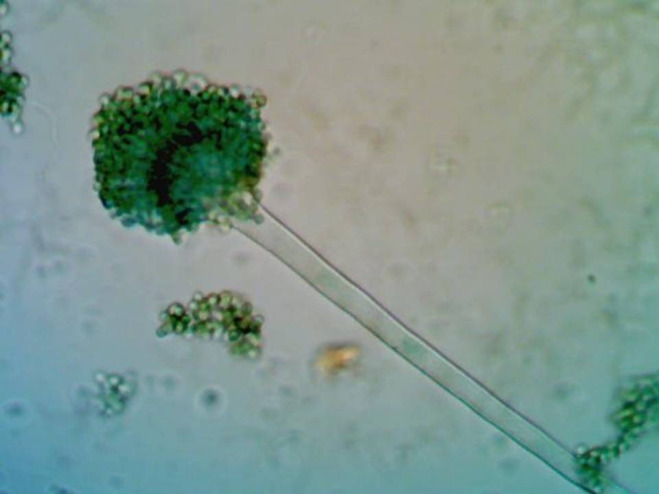
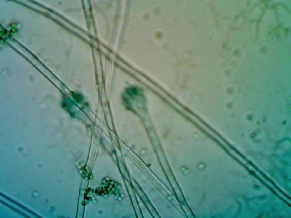
abdominal surgery and developed Aspergillosis. Colour on
SDA agar of Aspergillus fumigatus is olive green on
surface view and uncolored to cream on reverse view. The
colony texture is wooly. The outer edge of the colony is
white. The macro-micro culture and slide culture reveals
that the isolated pathogen is Aspergillus fumigatus.
A
B
Fig. 1 Aspergillus fumigatus (Photograph taken at 40X) (Slide culture)
CONCLUSION- Post-operative aspergillosis seems to be an underappreciated problem. Several reasons have been proposed for the increase in invasive fungal infections, including the use of anti-neoplastic and immunosuppressive agents, broad-spectrum antibiotics, and prosthetic devices and grafts, and more aggressive surgery. Invasive fungal infections in surgical patients are life threatening. The possibility of fungal infections should always be considered, especially in cases where the infection occurs a week or more after the surgery. Microbiological cultures of biologic fluids and tissue for the detection of an invasive fungal infection require multiple days and occasionally weeks for the identification of a specific fungal pathogen. In order to improve the outcome, better diagnostic methods are needed. Infected patients should receive during surgery. Prevention of these infections should include special care of the ventilation system in the operating theatre and adequate sterilization techniques.
ACKNOWLEDGMENT First of all we are thankful to Dr. Varsha Aglawe for her logistical support and for providing necessary guidance concerning work implementation and also grateful to Doctors and Hospital staff and patient for their cooperation.
REFERENCES
- Ahmad et al. (eds.). Combating Fungal Infections problems and remedy. Verlag Berlin Springer: 2010: pp. 22-45.
- Milana O, Aleksandra P, Nada V, Mlanda J. Mixed fungal infection (Aspergillus, Mucor and Candida) of severe hand injury. Hindawi,2014;2014
- Virat VH, Lebeau B, Bozonnet et al. severe filamentous fungal infections after widespread tissue damaged due to traumatic injury: six cases and review of the literature. Scandin avian journal of infectious diseases, 2009; 41: 491-500.
- Eggimann P, and Pitte D. Postoperative fungal infection. Surgical infection. 2006; 7:553-556.
- Prof. Detret kaya, et al. Fungal agents as a cause of wound infection. Wounds, 2007; 19(8):218-222.
- John HR, Michael AP, David W. Antifungal Susceptibility Testing: Practical Aspects and Current Challenges. Clin Microbiol, 2001; 14: 643–658.
- Gregory AE., Simon WL, Peggy LC. Antifungal prophylaxis in liver transplant recipients. J Liver transplantation, 2009; 15:842-858.
- Carlson GL, Mughal MM, Birch M, Denning DW. Aspergillus wound infection following laparostomy. J Infect, 1996; 33: 119–121.
- Pasqualotto AC, Denning DW. Post -operative Aspergillosis. Clin Microbiol infect, 2006; 12(11):1060-76.
- Olishola, Andrew Y, Zang, Curtin CM. Invasive Aspergillosis of the hand caused by Aspergillus ustus. J Hand, 2010; 5:102-105.
| International Journal of Life-Sciences Scientific Research (IJLSSR) Open Access Policy Authors/Contributors are responsible for originality, contents, correct references, and ethical issues. IJLSSR publishes all articles under Creative Commons Attribution- Non-Commercial 4.0 International License (CC BY-NC). https://creativecommons.org/licenses/by-nc/4.0/legalcode |
| How to cite this article: Tiwari M, Aglawe V, Arya P, Patel S, Jaget S: Case Report on Fungal Infection in Post Operative Patient. Int. J. Life. Sci. Scienti. Res., 2017; 3(1): 783-785. DOI:10.21276/ijlssr.2017.3.1.6 Source of Financial Support: Nil, Conflict of interest: Nil |