Key-Words- Dimethoate, Histopathology, Liver, Garra mullya
INTRODUCTION- Pesticides are occasionally used indiscriminately in large amounts causing environmental pollution and potential health hazards. Dimethoate is systemic insecticides produced by reacting salts of Dimethyldithio-phosphoric acid with N-methylchloroaecetamide, in aqueous medium in the presence of some organic solvents is widely used against a broad range of insects and mites and is also used for indoor control of houseflies. The extensive use of DM poses a health hazard to animals and humans because of its persistence in soil and crops [1]. One of the major agricultural chemical groups is pesticide which play important role in increasing agricultural productivity through controlling pest. But on the other hand, they cause much damage to the non-target organisms both in terrestrial and aquatic environment. Fish accumulate pollutants di-rectly from contaminated water and indirectly via food chain [2]. The runoff from treated areas enters the river and aquaculture ponds that are supplied by rivers and adversely affect the quality of water surfaces and creates hazards for aquatic life resulting in serious damage to non-target species, including fishes [3].Histopathology deals with the study of pathological changes induced in the microscopically structure of body tissue. Any alteration in normal structure of tissue indicates presence of disease or the effect of toxic substances like heavy metal and pesticides. [4] described histopathology as important tool for evaluating the action of any toxicant at tissue level. Histopathology provides data concerning tissue damage. Histopathological alterations can be used as indicators for the effect of various anthropogenic pollutants on organisms and are a reflection of overall health of the entire population in the ecosystem. The present study was under taken to analyze the impact of chronic concentration of dimethoate in liver of fish, Garra mullya.
MATERIALS AND METHODS- Healthy adult fish 4-5cm. in length Garra mullya were collected from local river Shivan Dist. Nandurbar, India. Fishes were washed with 0.1% of potassium permagnate (KmNo4) solution to avoid dermal infection. They were then rinsed in water and acclimatized to the laboratory conditions in the department of zoology for two weeks in 1000 l. capacity glass aquaria. Dead fish were removed immediately, Such as mortality may deplete dissolved oxygen with resultant effect on other fishes. During acclimatization fishes were fed with pieces of live earthworm on alternate days. Water also changed once in every day. The experiment was conducted natural and photoperiod of temperature 25Ģ1 ▒ 3Ģ20. Water quality was measure as per by [5], Conductivity- 0.64 ▒ 0.3, Dissolved O2- 6.3▒1.1 (ml/L), pH.- 8.60 ▒ 0.3, Acidity- 2.5▒ 0.1, Alkalinity- 44.1 ▒ 0.5, Total hardness- 67.5 ▒ 0.3. LC50 of dimethoate for 96 hours was determined by probit analysis method [6] (Finney, 1971). The animals were dissected and liver tissue carefully removed. Tissues were immediately washed in 1% saline solution to remove the adhering mucus and blood and soaked between the blotting papers. The tissue from the control and exposed batches were taken out and preserved in aqueous BouinÆs fluid for 24 to 48 hrs. This was followed by successive dehydration in ascending grades of alcohol. Tissues were cleared in xylene and embedded in paraffin wax (at 58░-60░C.). The tissue was then processed routinely and prepared into paraffin block cut at 6Ąm thickness using microtome and stained with Haematoxyline and Eosin [7]. Standard histo-pathological procedures were followed for histopathological investigations [8]. Observations were taken under light microscope.
RESULTS AND DISCUSSION - Histopathological changes have been widely used as biomarkers in the evaluation of the health of fish exposed to contaminants, both in the laboratory [9] and field studies [10]. In normal liver the hepatocytes form a cord-like pattern. These cords are arranged around tributaries of the hepatic vein. The liver cells are large in size, polygonal in shape with homogenous eosinophilic cytoplasm and centrally located nuclei. A large number of blood sinusoids are observed and separates the hepatic cords one from another. Exposure of dimethoate to Garra mullya induced histopathological changes in the liver. The hepatocytes have lost their normal architecture. The lumen of sinusoid contains mainly erythrocytes and macrophages. Numerous hepatocytes show marked cytoplasmic vacuolization. The liver cells are degenerated with necrosis because of lymphocytic infiltration. The histopathological changes of liver were more pronounced after the exposure period of fenvalerate [11], similar observation by [12]. Fish exposed to sub lethal concentration of dimethoate during 7, 14 and 21 days shown considerable degree of alteration in the liver.
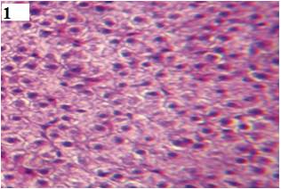
Fig. 1 normal structure of liver showing blood sinosoids and hepatic cells, centrally placed nucleus containing nuclei. (H&E, 450X)
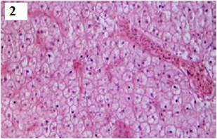
Fig. 2 On Dimethoate treatment after 7 days struc-ture of liver shows hemorrhage at some places vacuolation in the hepatic cells. (H&E, 450X)
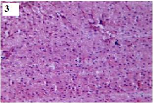
Fig. 3 On Dimethoate treatment after 14 days of liver shows haemorrhage at some places sinusoids are enlarged with vacuolation in the hepatic cells. Extensive degeneration of hepatocytes (H&E, 450X)
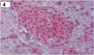
Fig. 4 On Dimethoate treatment after 21 days of liver shows extensive hemorrhage, necrotic hepatocytes, cloudy swelling and cytoplasmic vacuolization of hepatocytes (H&E, 450X)
The histopathological changes were more evident in specimens exposed to dimetoate and were not observed in the control fish. The liver cells in G.mullya are polygonal containing spherical central nucleus (Fig.1). After 7 days of exposure the hepatocytes became irregular and loose their polygonal shape. Some cells exhibited cloudy swelling, their contour becoming indistinguishable. There were many regions in the liver where cells were highly vacuolated. Many cells had exhibited pycnosis (Fig. 2 and 3). At the end of 21 days treatment, pronounced structural changes in hepatocytes such as focal necrosis, pycnosis, cloudy swelling and darkly stained specks of necrotic nuclei were observed (Fig.4). The majority of insecticides are bio-transformed in metabolites by liver through various enzyme systems and as a consequence of this process, liver undergoes different levels of damages. Alteration like irregular shaped hepatocytes, cytoplasmic vacuolation and laterally placed nuclei were also observed in the siluriform Corydoras paleatus after exposure to organophosphate pesticide for 96 hrs observed by [13]. [14] also observed lipid va-cuoles, hepatocytes swelling and pyknotic nuclei in Oreochromic nilotitus exposure to alachlor for 96 hrs. [15] also observes that dimethoate is strongly hepatotoxic and severely affect histology, carbohydrate and protein metabolism on liver of fish, Cyprinus carpio. [16] reported histopathological lesions induced in the hepatopancreas of Channa punctatus and Clarius batrachus exposed to industrial pollutants.
CONCLUSION- The present investigation shows After 7, 14 and 21 days of exposure of Dimethoate the hepatocytes became irregular, cloudy swelling and loose their polygonal shape. Liver cells were highly vacuolated, pycnosis and necrotic nuclei were observed in the histological structure of liver in Garra mullya. The changes in liver are biomarker in the evaluation of health of fish. The metabolic activities of the fish are affected which in turn become lethal to the fish. Dimethoate used to protect many field crops against disease, hence farmer come direct contact it and may affect their health.
REFERENCES
- WHO/IPCS (1996) Principles and methods for assessing direct immune toxicity associated with exposure to chemicals. Environ. Health Criteria 180: 110-112.
- Sasaki, Y., Izumiyama, F., Nishidate, E., Ishibashi, S., Tsuda, S., Matsusaka, N., Asano, N., Saotome, K., Sofuni, T. and Hayashi, M. (1997) Detection of genotoxicity of polluted sea water using shellfish and the alkaline single-cell gel electrophoresis (SCE) assay: A preliminary study.Mutation. Res. 393; 133-139.
- Bondarenko, S., Gan, J., Haver D.L. and Kabashima, J.N. (2004) Persistence of selected organophosphate and carbamate insecticides in waters from a coastal watershed, Environmen. Toxicol. and chemistry, 23(11); 2649-2654.
- Sprague J.B. (1973) The ABCÆs of pollutant bioassay using fish. In biological methods for the assessment of water quality, Am. Soc. Test. Mater. Tech. Publ., 528, 6-36.
- APHA AWWA and WPCF (2005) Standard methods for the examination of water and wastewater. 21st Edn.Washington DC USA.
- Finney, D.J., (1971) æProbit analysisÆ Third edition, Cambridge University, Press.
- Luna L.C (1968), manual of histologic staining methods. Armed Forces Institute of Pathology, 3 rd Ed. McGraw Hill Book Company, New York.
- Robert, R.J. (1989) Fish pathology (2Ænd edition), London, Billier, Tindall. 11, pp 453.
- Thophon, S., Kruatrachue, M., Upathan, E.S., Pokethitiyook, P., Sahaphong, S. and Jarikhuan S. (2003) Histopathological alteraions of white seabass Lates calcarifer in acute and subchronic. Environ. Poll., 121: 307-320.
- Teh, S. J., Adams, S.M. and Hinton, D.E. (1997) Histopathological biomarkers in feral fresh water fish populations exposed to different types of contaminant stress. Aquatic Toxicol., 37:51-70.
- Sakr, S. A. and Jamal Al lail, S.M. 2005. Fenvalerate Induced Histopathological and Histochemical changes in the Liver of the Cat fish Clarius Gariepinus. Egypt. J. Aquatic Biol. Fish.,6 (2):103-124.
- Sridhar, K. and Esther Joice, P. (2012) Carbendazim induced histopathological and histochemical changes in liver tissue of common carp, Cyprinus carpio. Inter. J. of Life Sci. 5(1); 65-70.
- Fanta, E,F. Ritos, S., Ramao, S. Vinna, A.C.C. and Friberger, S. (2003) Histopathology of the fish, Corydoras paleatus contaminated sub lethal of organophosprus in water and food. Ecotoxicol. Environ. Saf., 54;119-130.
- Peebua, P.M., Kruatrachue, Pokethitiyook P. and Singhakaew, S.(2008) Histopathological alteration in Nile thilapia, Oreochromic nilotitus in acute and sub chronic alachlor exposure. J. of Environ. Biology, 29, 325-331.
- Ram Narayan Sing. (2013) Effect of dimethoate (30% EC), an organophosphate pesticide on liver of common carp, Cyprinus carpio, J. of Environ. Biology. 34,657-661.
- Bhattacharya, T., Ray, A.K. and Bhattacharya, S. (1984) Histopathological lesions in the haepatopancreas of Channa punctatus (Bloch.) exposed to mixture of mercuric chloride, phenol and a factory effluent. Matsya.11:1.
| Source of Financial Support: UGC (WRO) Pune Conflict of interest: Nil |
Ā
| International Journal of Life-Sciences Scientific Research (IJLSSR) Open Access Policy Authors/Contributors are responsible for originality, contents, correct references, and ethical issues. IJLSSR publishes all articles under Creative Commons Attribution- Non-Commercial 4.0 International License (CC BY-NC). https://creativecommons.org/licenses/by-nc/4.0/legalcode |
Ā