OBJECTIVE- To assess the incidence of different shapes of the medial and lateral meniscus and the incidence of discoid meniscus in the North Indian population.
METHODS- The study included 112 menisci from 56 adult cadavers knee joint irrespective of sex that were preserved in 10% formalin. After methodical dissection procedure the morphological variants of the shape of menisci were macroscopically noted and classified. The medial menisci were sub-grouped as crescent-shaped, V- shaped, U- shaped, C- shaped and sickle shaped. Lateral menisci were sub-grouped as crescent-shaped, C- shaped and discoid-shaped.
RESULTS AND CONCLUSIONS- In the present study, 80% of the medial menisci were crescent shaped, 12.8% were V-shaped while 7.2% sickle-shaped. Among the lateral menisci, maximum number of specimen were C-shaped i.e. 78% followed by crescent shaped in 19% and discoid shaped in 3% specimen. Knowledge of various shapes of meniscii would be useful for health professionals who treat athletes with suspected meniscal tear.
Key-Words- Knee, Lateral meniscus, Medial meniscus, Shape, Discoid
INTRODUCTION- The Menisci of the knee joint are to be considered main elements for perfect articulation among the articular osseous surfaces. Menisci are semicircular shaped fibro cartilagenous structures with bony attachments at the anterior and posterior aspect of tibial plateau and are wedged between femoral condyles and tibial plateau, the medial and lateral sides of the knee joint. [1] It is a double condyloid joint with 20 degree of freedom of motion. The functions of menisci include shock absorption, load transmission and improve joint stability pro-prioception, joint lubrication and nutrition. [2] The anatomical abnormalities and variation of the intra-articular struc-tures of knee joint have recently gain importance because of new techniques such as arthoscopy, com-puted tomography and magnetic resonance imaging investigation of these va-riants are important in order to define the morphological features for surgical procedures and clinical diagnosis. [3] The menisci have several roles that contribute to the successful function of the knee. Injuries to the meniscus are common in activities and sports. Long term complication of removal of a meniscus includes cartilage degeneration and bone remodeling. [4] Hence, today a ruptured meniscus is repaired rather than removed, but this treatment is only feasible when the meniscus is of good quality. [5] Hence, this study was undertaken with the objective to estimate the incidence of different shapes of medial and lateral meniscus and also the incidence of discoid meniscus in North Indian population.
MATERIALS AND METHODS- The study was conducted over a period of two years in the department of Anatomy, Katihar Medical College Katihar, Bihar, India. For this study, human adult knee joint available in the anatomy laboratory were used. The study included 112 menisci from 56 knee joints of the North Indian population. All specimens preserved in a solution of 10% formalin were used. Menisci which showed any structural change due to injuries or advanced degenerative changes were excluded as that may prevent its morphological analysis. After the dissection of skin and muscles, the approaches to the menisci were performed, opening anteriorly by a longitudinal incision on each side of the joint capsule, cutting the patellar ligament and the collateral ligaments transversely to expose the menisci clearly, the joint capsule and the intra-articular ligaments were cut and the condyles were circumferentially detached from their soft tissue attachments and removed, exposing the tibial plateau. The dissection procedures were performed in a systematic fashion and the data were recorded on a standardized collection sheet.
Morphological variants of the shapes of the meniscus were macroscopically noted and classified. The medial meniscus (MM) was sub-grouped as crescent shaped, U-shaped, V-shaped, C- shaped and sickle shaped. The lateral meniscus (LM) was sub-grouped as crescent (semilunar) shaped, C- shaped and discoid shaped. When the meniscus covers the tibial plateau circularly the meniscus is said to be discoid. The incomplete discoid menisci had an opened area at the centre of menisci and they were horse shoe shaped. The menisci which did not have any opened area at the centre of the menisci were defined as complete discoid menisci.
Menisci which had the anterior and posterior horns and a thin body were defined as crescent (semilunar) type. The meniscus which had thin anterior and posterior horns and a thick body were defined as sickle shaped type.
RESULTS- Study was done on 112 menisci. It was observed that 80%of medial menisci were crescent shaped, 12.8% showed V-shaped and 7.2% were sickle shaped. Among the lateral menisci 78% were C-shaped, 19% were crescent shaped and 3% showed incomplete discoid.
Table: 1 Showing the incidence of different shapes of medial meniscus (n=56)
| Shape | Total & Percentage (%) | á
|---|---|
| Crescentic | 45 (80%) |
| Sided V | 07 (12.8%) |
| Sickle shaped | 04 (7.2%) |
Table: 2 Showing the incidence of different shapes of lateral meniscus (n=56)
| Shape | Total & Percentage (%) | á
|---|---|
| C shaped | 44 (78%) |
| Crescentic | 10 (19%) |
| Discoid | 02 (3%) |
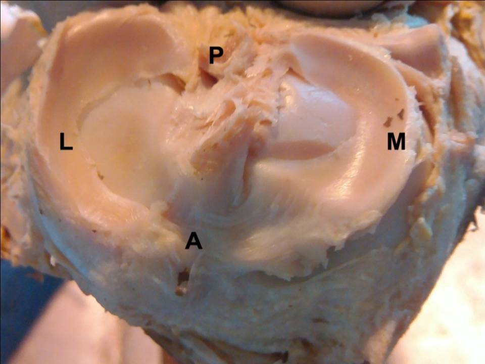
Fig 1: Right tibial plateau showing C-shaped lateral meniscus
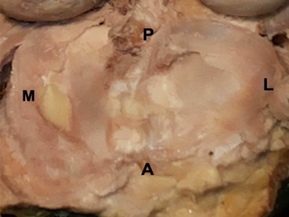
Fig 2: Incomplete discoid lateral meniscus of left knee joint
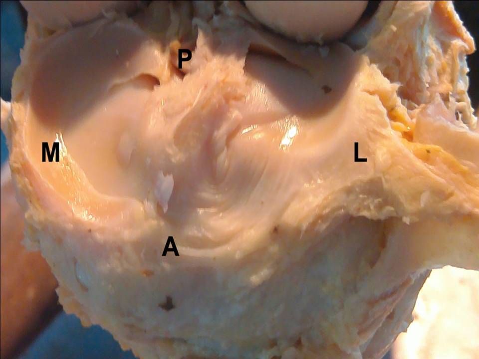
Fig 3: Left tibial plateau showing cresent shaped medial meniscus
In primates, Parsons noted that medial meniscus (MM) always has a crescentic shape but the lateral meniscus (LM) may have either a crescent or disc shape. Vallois [6] confirmed the observation and studied the whole morphology of the knee joint in primates.
Flick & Rudolph [7] described the medial meniscus (MM) as a half, two third or three-fourth ellipse and stated that the lateral could be compared to an almost complete circle. In contrast, Charles [8] classified the menisci, both on the basis of the relative size of the anterior and posterior cornua and also on the degree of curvature. In 1889, Young [9] described a discoid lateral meniscus in a cadaver specimen. Discoid meniscus is an atavistic anomaly in which the meniscus of the knee, predominantly LM, is discoid rather than semilunar in shape. [10] The fact was mentioned as the discoid meniscus was most likely a congenital deviation and usually occurred laterally. Moreover it was reported that the discoid shape resulted in a greater coverage of the tibia and was usually associated with increased thickness of the meniscus that might lead to abnormal shearing forces across the knee joint [11].
The most common congenital abnormality of the me-niscus in man is a discoid meniscus with a reported incidence of 0.4 to 17%, the vast majority occurring on the lateral side of the knee. [12] From a study conducted by Rao and Rao [13] in south India based on 3167 knee arthroscopies done between the years 1993 and 2004, 177 (5.59%) discoid lateral menisci were encountered. The present study re-ported the incidence of discoid meniscus 3% from the North Indian population. Our result that in majority of specimen (80%), the medial meniscus (MM) was crescent shaped and the most common shape of lateral meniscus (LM) was C-shaped (78%). The study has provided information on different shapes of lateral and medial meniscus with contribution to a better delineation of meniscal anatomy.
CONCLUSION- From our study we can conclude that in most of the specimen the medial meniscus was crescentic in shaped (80%). Commonest incidence of lateral meniscus was C-Shaped (78%). Incomplete lateral discoid menisci were observed in 3% of lateral meniscus. Our study will provide support to the meniscal anatomy concerning the surgical procedures and of the knee joint. The study has provided further information on different shapes of the medial and lateral meniscus especially the presence of incomplete lateral discoid menisci in adults which is a more important finding. This study is useful for the health professional who work with the treatment of meniscal injuries to create an awareness of the anatomical variation that exist in the menisci facilitating the rehabilitation process.
ACKNOWLEDGMENT- We thank to Prof N. K. Pandey and all the staff of department of Anatomy KMC Katihar for their invalua-ble advice and support. The authors are grateful to au-thors/ editos/ publishers of all those articles, journals and books from where the literature of this article has been reviewed and discussed.
REFERENCES
- [1] Buckwalter JA, Amendolam A, Clark CR. Articular cartilage and meniscus. In: Insall and Scott NW. Surgery of the knee. 1st ed. London: Churchill Livingstone; 1986.p.310.
- Gray JC. Neural and vascular anatomy of the menisci of the human knee. J Orthop Sports Phys Ther. 1999; 29 (1): 23-30.
- Moore KL, Dalley AF. Clinically oriented anatomy. 4th ed. Philadelphia: Lippincott Williams and Wilkins; 1999. P 690-99.
- Fairbank TJ. Knee joint changes after meniscectomy. Journal of Bone and Joint Surgery 1948; 30B:664-70.
- Messner K, Gao J. The menisci of the knee joint: Anatomical and functional characteristics and a rationale for clinical treatment. J Anat 1998; 193: 161-78.
- Vallois H. Etude Anatomique de, Articulation du Genou chez les Primates. Montpellier: Lĺabeille. 1914
- Fick & Rudolph. In: Bardelebenĺs Handbuch der Anatomie des Menschen: II Band, Handbuch der Anatomie und Mechanik der Gelenke, Teil I und III, S. G. Fischer, Jena. 1904; 354-358.
- Charles CM. On the menisci of the knee joint in American Whites and Negroes. Anat Rec 1935; 63:355-364.
- Young R. The external semilunar cartilage as a complete disc. In: Cleland J, Young R, eds. Memoris and Memoranda in Anatomy. London: Williams and Norgate, 1889; 179.
- Parson HG. The external semilunar cartilage of the knee in the primates. J Anat 1900; 34:32.
- Kelly BT, Green DW. Discoid lateral meniscus in children. Curr Opin Pediatr 2002; 14: 54-61.
- Dickhaut SC, DeeLee JC. The discoid lateral meniscus syndrome. J Bone Joint Surg Am 1982; 64: 1068-1073.
- Rao SK, Rao PS. Clinical, radiologic and arthroscopic assessment and treatment of bilateral discoid lateral meniscus. Knee Surg Sports Traumatol Arthrosc 2007; 15: 597-601.
| Source of Financial Support: Nil Conflict of interest: Nil |
á
|---|
| International Journal of Life-Sciences Scientific Research (IJLSSR) Open Access Policy Authors/Contributors are responsible for originality, contents, correct references, and ethical issues. IJLSSR publishes all articles under Creative Commons Attribution- Non-Commercial 4.0 International License (CC BY-NC). https://creativecommons.org/licenses/by-nc/4.0/ |
á
|---|