SSR Inst. Int. J. Life Sci., 9(1):
3147-3150,
January 20223
Mucometra due to
Follicular Cyst in an Ongole Cow- A Case Report
Anthony Raju Kangonu1*, Chandra
Prasad Borra2, Srinivas Manda3
1PG
Scholar, Department of Veterinary Gynaecology and
Obstetrics, NTR College of Veterinary Science, Gannavaram,
Sri Venkateswara Veterinary University, Tirupati, India
2Assistant
Professor, Department of Veterinary Clinical Complex, NTR College of Veterinary
Science, Gannavaram, Sri Venkateswara Veterinary
University, Tirupati, India
3Professor,
Department of Veterinary Gynaecology and Obstetrics,
NTR College of Veterinary Science, Gannavaram, Sri
Venkateswara Veterinary University, Tirupati, India
*Address for Correspondence: Dr. Kangonu Anthony Raju, PG
scholar, Department of Veterinary Gynaecology and
Obstetrics, NTR college of veterinary science Gannavaram,
Sri Venkateswara veterinary university, Tirupati, India
E-mail: vetanthony1693@gmail.com
ABSTRACT- Background: An eight-year-old Ongole
cow was brought to the large Gynaecology ward,
Department of VGO, NTR College of Veterinary Science, Gannavaram
with a history of irregular cloudy vaginal discharge. The local veterinarian
did not appreciate the growth of the gravid uterine horn during repeated
per-rectal examinations in 30-day intervals.
Methods: On rectal examination, the right uterine horn was distended with fluid.
On real-time ultrasonography, the ovaries were diagnosed with the presence of
large anechoic follicles on both left and right ovaries. The cow was diagnosed
as mucometra due to follicular cyst and treated with ovsynch
plus CIDR protocol using 20µg of GnRH and cloprostenol
sodium of 500 µg and CIDR device containing progesterone of 1.9 gms.
Results: Re-examination after one month revealed the persistence of cysts
on both the ovaries and the distended right uterine horn.
Conclusion: The treatment was not successful because of the longstanding
follicular cysts and thickening, and unresponsiveness of uterine endometrium.
The prognosis of the present case was guarded.
Key Words: Follicular cyst, Mucometra, Ongole cow, Ovsynch plus
CIDR, Ultrasonography
INTRODUCTION- Cystic ovarian (OC) condition
is the important cause of infertility in milch cattle and is defined as enlarged
anovulatory follicle-like structures persisting for 10 or more days in dairy
cows. Nowadays it is explained as
follicular structures that are present on the ovaries with a diameter of not
less than 17 mm for more than 6 days in the absence of CL [1]. A cystic follicle can persist as a dominant structure effectively
preventing follicular growth and can be replaced by another cystic follicle or
regress. [2]. Long-term continuance of
follicular cysts leads to hypertrophy of the endometrial glands, resulting in
mucometra [3]. Failure to ovulate leads to cyst development,
interfering with normal ovarian function. Grossly, Ovarian cysts are of two
types, follicular cysts, and luteal cysts. These cysts can be discerned by
examining progesterone concentration in milk and blood plasma. Ultrasound
examination of wall thickness can be useful in differentiating these cysts
[4]. The oestrous cycle is not blocked by the
cystic condition of the ovaries, which is frequently accompanied by other
alterations in the ovaries and by damaged endometrium. Follicular cystic
condition showed unusual subepithelial layer density in the uterus [5].
The hallmark of mucometra or hydrometra is the build-up of mucin like substance
in the uterus. Mucometra is frequently linked to higher progesterone
stimulation in ovine and caprine, but in cows, mares, and bitches, it is due to
increased progesterone or oestrogen stimulation [6].
The echogenicity of the uterine content is the Eco graphical distinction
between mucometra and pyometra. Unlike sterile mucus, which appears anechoic,
purulent mucus exhibits some degree of echogenicity [7]. However, in
the present report, the consistency and mucoid content of the uterine fluid
prompted a diagnosis of mucometra. The mucometra was accompanied by endometrial
hyperplasia and dilation of the endometrial glands, which can be concluded to
be caused by persistent follicular cysts.
CASE PRESENTATION- An eight-year-old pluriparous Ongole cow was
presented to the Department of VGO, Gannavaram,
Krishna district with a history of irregular cloudy discharges and a calving
history of 2 years and artificial insemination was performed six months ago.
The local vet did not notice any uterine horn enlargement even after repeated
rectal examinations in the 30-day interval. The clinical parameters
(temperature, pulse, and heart rate) were within normal range. Physical
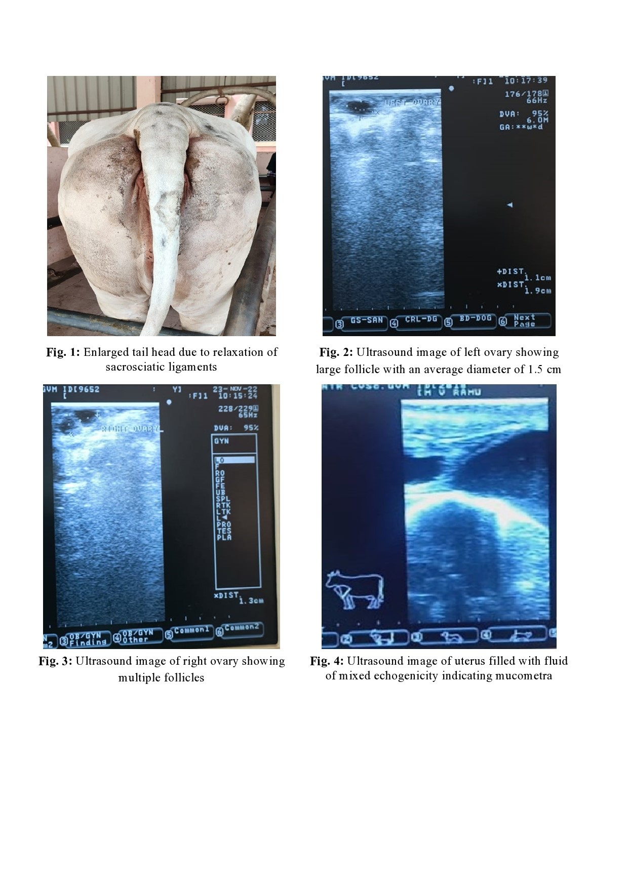
examination of the animal revealed bull-like appearance (masculine physical
traits) of the cow and an enlarged tail head was noticed due to the relaxation
of sacrosciatic ligaments (Fig. 1). Per-rectal examination, revealed enlargement
of the right uterine horn and distended with the absence of foetal
membrane slip and fremitus. Ultrasonography of the uterus and ovaries revealed
the presence of cystic follicles with an average diameter of 1.5cm on the left
ovary (Fig. 2) and multiple follicles on right ovary (Fig.
3) with a fluid of mixed echogenicity accumulated in the lumen of the uterus
(Fig. 4).
Based on the findings the
case was diagnosed as mucometra due to persistent follicular cysts.
TREATMENT- The
present case was treated with Ovsynch plus CIDR
protocol. The animal was given an intramuscular injection of 20 µg GnRH (Pregulate, 4 µg/ml) on the 0th day, insertion of a CIDR device containing
progesterone of 1.9 gms on the 0th Day,
500 µg of cloprostenol sodium (Pragma 250 µg/ml)
intramuscularly along with CIDR removal on 7th day and 20 µg of GnRH
(Pregulate, 4 µg/ml) on 9th day. A total
10 gm of KI was given per oral for 5 days and Repronol
(vitamin E and Se) at a dose of 5 ml was given twice intramuscularly within 10
days. Transrectal ultrasonography was done one month after the treatment, and
the follicles on both the right and left ovary remains unchanged.
DISCUSSION- However,
in the present case, the consistency and mucoid content of the uterine fluid
prompted a diagnosis of mucometra. Treatment of follicular cysts with
progesterone impedes the endocrine environment required to maintain the
follicular cysts and results in the restoration of ovarian cyclical activity [1].
Ultrasound-guided ablation is a safer method, which avoids adhesion and
bleeding in the ovary. The estrogens produced by the follicular cysts have a
preventive effect on ovulation. Therefore, ablation of the cyst will destroy
the estrogen source, leading to new follicular waves and ovulation [8].
The mucometra was accompanied by endometrial hyperplasia and dilation of the
endometrial glands, which can be concluded to be caused by persistent
follicular cysts. A similar case was also reported by [9], in which
mucometra was associated with follicular cysts.
Follicular cysts can be treated with GnRH, which
causes the release of luteinizing hormone (LH) and luteinization of the cyst.
The luteinised cyst can be sensitive to PGF2α, and regress about 8-9 days later with the
administration of PGF2α [10]. In the present case, an attempt
was made to bring about ovulation by intramuscular injection of GnRH. However,
the follicles on both the left and right ovary are persistent.
Circulating
progesterone levels are enhanced with the treatment using a CIDR device is
effective in rectifying follicular cyst conditions [11]. Exposure of
exogenous progesterone to cows with unresponsive hypothalamus restores the
ability of E2 to induce the release of LH in a surge-like manner [12,13]. In the present case, this line of treatment
was not attempted due to the thickening of the endometrium and oedema of the
endometrial glands, which would be unable to respond to gonadotropin
stimulation.
CONCLUSIONS-
Mucometra may be confused as early gestation, but it can be discerned by the
absence of fetal membrane slip and ultrasonic examination of the reproductive
tract. The present case report is mucometra due to follicular cyst, its
diagnosis and management. The ACTH hormone released, because of stress causes
increased levels of progesterone, at sub luteal dose even after the luteolysis for several days leading to the formation of
persistent follicles. Nowadays confinement of an animal in its shed for longer
periods without any exercise leads to a lot of stress for animals. However, the
treatment for the present case was not successful due to the persistence of the
cysts on the ovaries for longer periods and damage to the endometrial glands.
The prognosis of the present case was grave.
The follicular cysts occurred after postpartum and
eventually rebound to normal ovarian activity if the proper diagnosis was made
at the early stages. Misdiagnosis at the early stages of cystic condition leads
to mucometra or hydrometra resulting in worsening the reproductive potential of
the animal. The etiological factors like Stress full conditions to the animal,
lack of exercise, and high protein diet should be corrected to bring about good
results.
CONTRIBUTION OF AUTHORS
Research concept- Manda Srinivas
Research design- Manda Srinivas
Supervision- Borra Chandra Prasad
Materials- Kangonu Anthony Raju
Data collection- Kangonu Anthony
Raju
Data analysis and Interpretation- Kangonu Anthony Raju
Literature search- Kangonu Anthony Raju
Writing article- Kangonu Anthony Raju
Critical review- Borra Chandra Prasad
Article editing- Borra Chandra Prasad
Final
approval-
Manda Srinivas
REFERENCES